

Researchers at The University of Texas MD Anderson Cancer Center have developed a spatial map of muscle-invasive bladder cancer, revealing how tumor cell states, immune environments and therapeutic vulnerabilities are organized within tumors. The study, published in Cancer Discovery, provides a new framework for understanding why patients with bladder cancer may respond differently to treatment.
The research was led by Linghua Wang, M.D., Ph.D., professor of Genomic Medicine, executive director and head of the Center for Cellular Language Intelligence, associate member of the James P. Allison Institute™, and focus area co-lead with the Institute for Data Science in Oncology; together with Jianjun Gao, M.D., Ph.D., professor of Genitourinary Medical Oncology, and co-first authors Kai Yu, Ph.D., postdoctoral fellow in the Wang laboratory, and Jianfeng Chen, M.D., Ph.D., instructor of Genitourinary Medical Oncology.
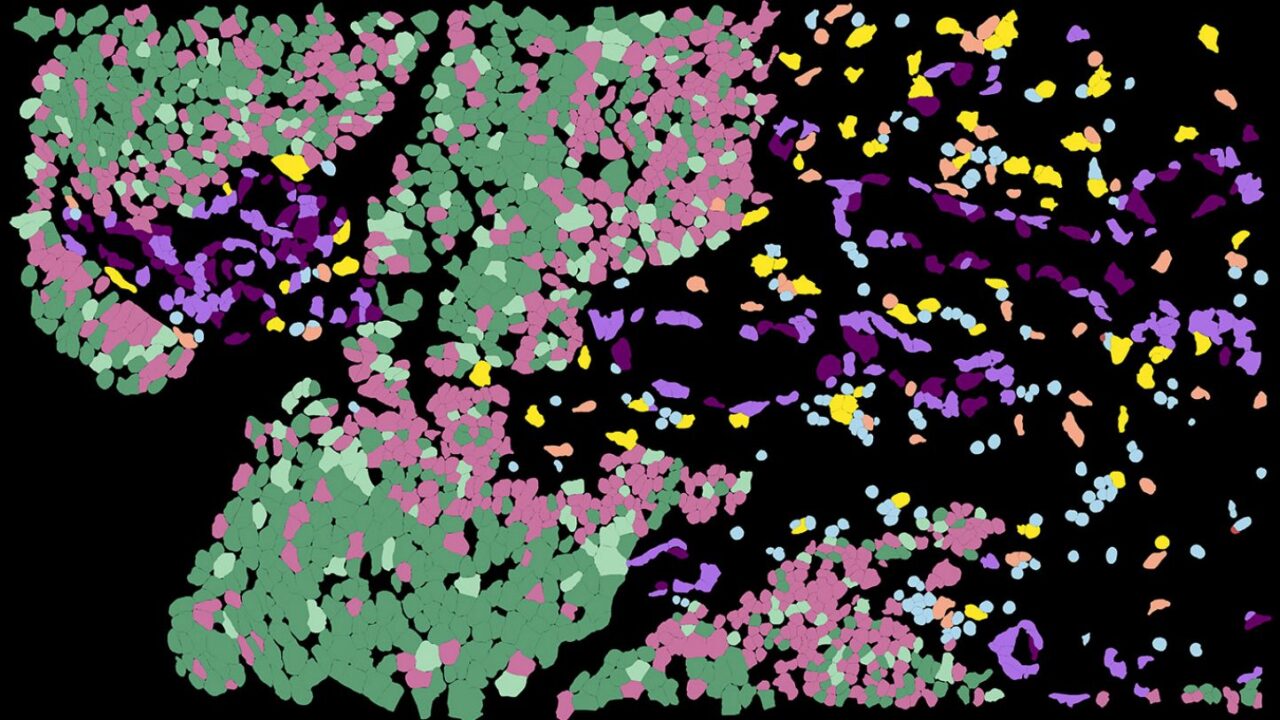
“Traditional molecular subtyping often classifies bladder cancers as either luminal or basal, but our spatial analyses show that this binary view is incomplete,” Wang said. “Within a single patient’s tumor, luminal and basal-like programs can coexist in highly organized spatial patterns, and those patterns are closely tied to immune activity, lineage-specific treatment vulnerabilities, and how different tumor regions may respond to treatment.”
The study supports the development of spatially informed biomarkers that could help researchers and clinicians better understand which therapies are most likely to affect specific tumor regions and tumor cell states. For example, future approaches could evaluate NECTIN4-directed therapies for immune-cold luminal tumor cores, while using chemotherapy or immunotherapy-based treatments may be a better strategy to target inflamed basal-like margins.
Future studies will need to validate these findings in larger, prospective clinical cohorts, including post-treatment samples, to determine how therapy reshapes tumor architecture over time.