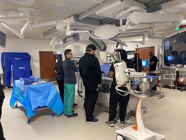
Doctors at Houston Methodist Neuroscience & Spine Center at Sugar Land continue to advance neurological care with new technologies that improve patient treatment and safety.
“We are seeing rising numbers of stroke patients across the country, due to a variety of factors,” said Tsz Lau, M.D., a board-certified neurosurgeon at Houston Methodist Sugar Land Hospital. “We need to be able to treat more patients, faster, to ensure that we are protecting brain function and providing patients with the best possible quality of life post-treatment. That means restoring blood flow to the brain as quickly as we can. Innovative technology gives us a significant advantage both in terms of better visualization of the vascular system and brain and the ability to make rapid adjustments in the new neuro lab.”
The hospital recently installed an advanced image-guided therapy system for neurological conditions that enhances patient safety and improves the visibility of the entire brain.
“This new system provides excellent visibility and precise views at different angles and with various patient anatomy,” said Lau. “This is especially critical in stroke care, where time is so important. Because of the quality of the images produced, we can perform procedures more efficiently, which can reduce treatment time by as much as 30 minutes or more.”
The new image-guided technology allows neurosurgeons to switch smoothly between 2-D and 3-D imaging during stroke treatment and gives them the ability to treat more complex cases.
“This is a big step forward for us,” said Lau, medical director of cerebrovascular surgery. “Houston Methodist Sugar Land Hospital is committed to investing in the latest technology and it makes a significant difference in the results we can achieve for our patients. We experienced this recently when we completed the first minimally invasive surgery to remove a blood clot in the brain, also known as a thrombectomy, in Fort Bend County.”
The FDA-approved optical imaging agent is used during fluorescence-guided surgery (FGS) in patients with high-grade glioma. The patient takes the drug orally several hours before surgery, which allows the neurosurgeon to view the brain through a surgical microscope outfitted with special blue filters. The drug makes cancerous cells glow a red-violet color under the filter, which makes it easier to distinguish them from healthy cells.
“One of the major challenges in removing a brain tumor is protecting as much healthy tissue as possible to preserve critical functions such as speech or balance,” said Lau. “With this new imaging agent, I can get a much clearer picture of where the tumor begins and ends, which allows me to operate with much higher levels of precision.”


